







Images from Inside the Body
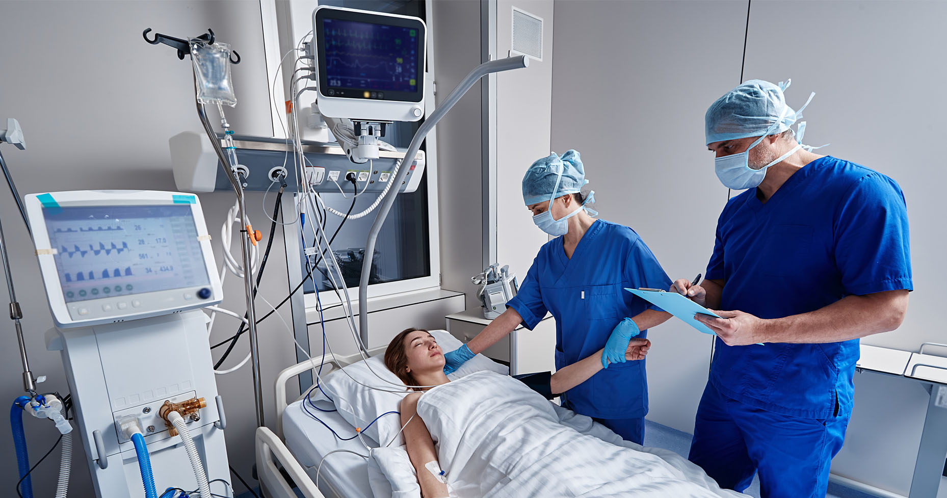
The radiology department is a central unit that works in close cooperation with all branches in our hospital. In many cases, a reliable diagnosis can only be reached after radiological imaging. For our radiology team, an individual approach to the patient and high-precision diagnosis are a priority. Our goal is to provide the most appropriate imaging with the lowest possible radiation dose and to carry out the examination at the highest quality and deliver the results to the physicians as soon as possible.
Why Çakırtepe?
Extensive Professional Experience
Our radiology team has many years of experience in the field of imaging and works in collaboration with university hospitals when necessary.
Service in Emergencies
Our radiology unit provides 24/7 service to provide rapid imaging and diagnosis in emergencies.
Choosing the Right Method
Imaging methods such as Computed Tomography (CT), Magnetic Resonance (MRI), Ultrasound, X-ray, and Mammography are available in our hospital. The most appropriate method is selected according to the patient's condition.
Ultrasound
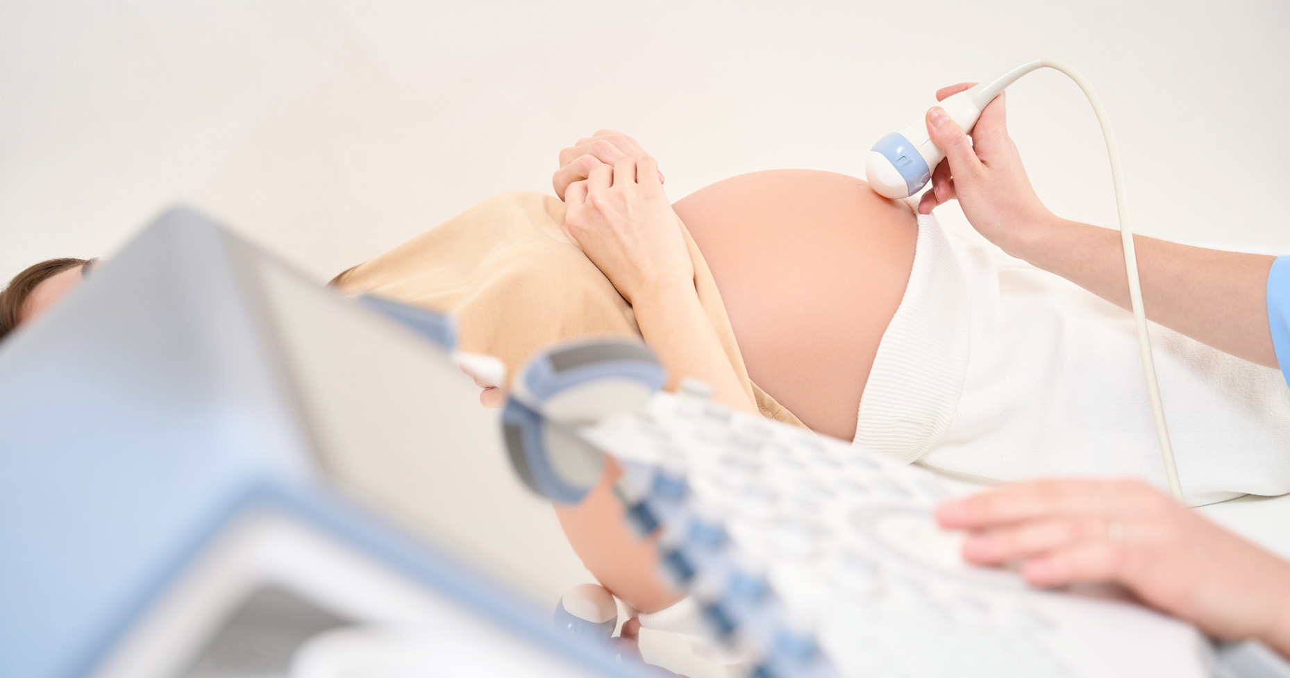
Ultrasound (sonography) is a radiation-free imaging method that works with sound waves. Organs such as the liver, kidneys, and spleen, which cannot be imaged adequately with an X-ray, can be evaluated clearly with ultrasound. In addition, ultrasound-guided tissue sampling (puncture) or drainage of fluid accumulations can be performed.
Areas of Use of Ultrasound
- Pregnancy follow-up
- Acute abdominal diseases (e.g., gallstones or kidney stones)
- Evaluation of vessels (arteries and veins) and examination of their patency
- Examination of the thyroid gland, heart, kidneys, and bladder
Mammography
Mammography is an imaging method that allows the breast tissue to be examined by X-ray method and is used in the early diagnosis of tumors. With the early detection of breast cancer, treatment success is highly possible. Our experienced radiology specialists accompany you during the examination process.
Indications for Mammography
- For screening purposes in women over 40
- In the presence of a palpable mass in the breast
- When hardness is felt in the breast tissue or a part of it
- When there are palpable enlarged lymph nodes under the armpit
- In high-risk situations due to family history or previous breast cancer diagnosis
X-ray
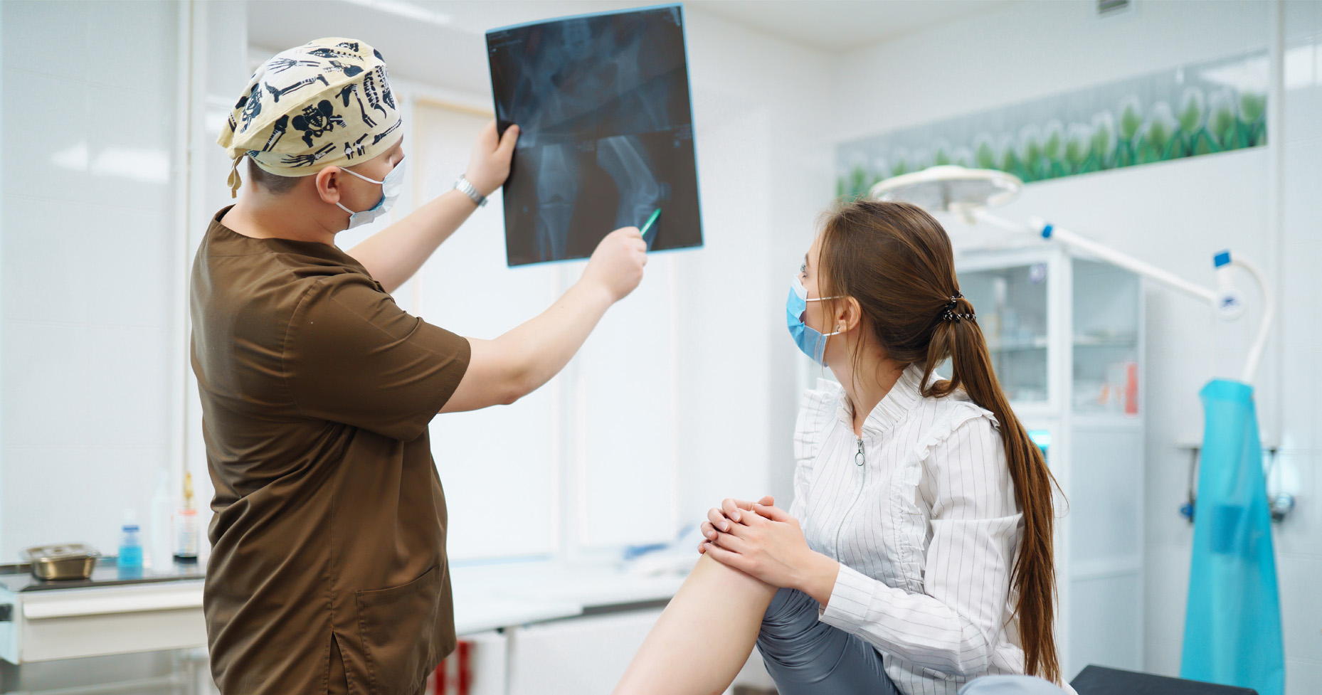
The X-ray device provides imaging of structures in the skeletal system, heart, lungs, and abdomen. Images are instantly reflected on the screen and a quick evaluation can be made.
Areas of Use of X-ray
- Thorax (chest)
- Bones and joints
- Evaluation of joint movements
- Detailed three-dimensional imaging in fine bone fractures
- Detailed and fast imaging of brain, neck, abdominal, and peripheral vessels with CT Angiography
Computed Tomography (CT)
With computed tomography, cross-sectional images of the body are obtained in very high resolution. Contrast material may be administered orally or intravenously during the examination. The procedure is painless and images are instantly evaluated on the computer screen. In this way, the necessary treatment can be started quickly.
Areas of Use of CT
- Head region complaints (brain hemorrhage, stroke)
- Lung diseases (infections, pulmonary embolism, tumors)
- Intra-abdominal organ and intestinal diseases
- Evaluation after severe traumas
- Tumor staging
Magnetic Resonance Imaging (MRI)
Magnetic resonance imaging uses magnetic fields and radio waves to examine organs as well as muscles, tendons, and ligament structures in detail. Cross-sectional images of the examined area are obtained.
Areas of Use of MRI
- Brain
- Joints (shoulder and knee)
- Vascular structures, MR angiography
- Intra-abdominal organs
- Soft tissues (muscles and tendons)
Bone Densitometry (DEXA)
Bone densitometry is a reliable low-dose radiation imaging method that measures bone mineral density to assess bone health. It is particularly used in the early diagnosis of osteoporosis and the procedure is performed quickly and painlessly.
Areas of Use of Bone Densitometry
- Diagnosis and follow-up of osteoporosis (bone loss)
- Evaluation of fracture risk
- Post-menopausal bone density follow-up
- Monitoring of patients with long-term cortisone use
- Evaluation of treatment effectiveness in diseases affecting bone health
Department Doctors

Dr. Serdar Altay
Radiology